
Find your area of care.
Select an area to explore its procedures. Laparoscopy is an approach that can be used for different operations.
General surgeryGallbladder, hernias, digestive system and soft tissues.
Gallbladder, liver and bile ducts
- Gallbladder removal (cholecystectomy)
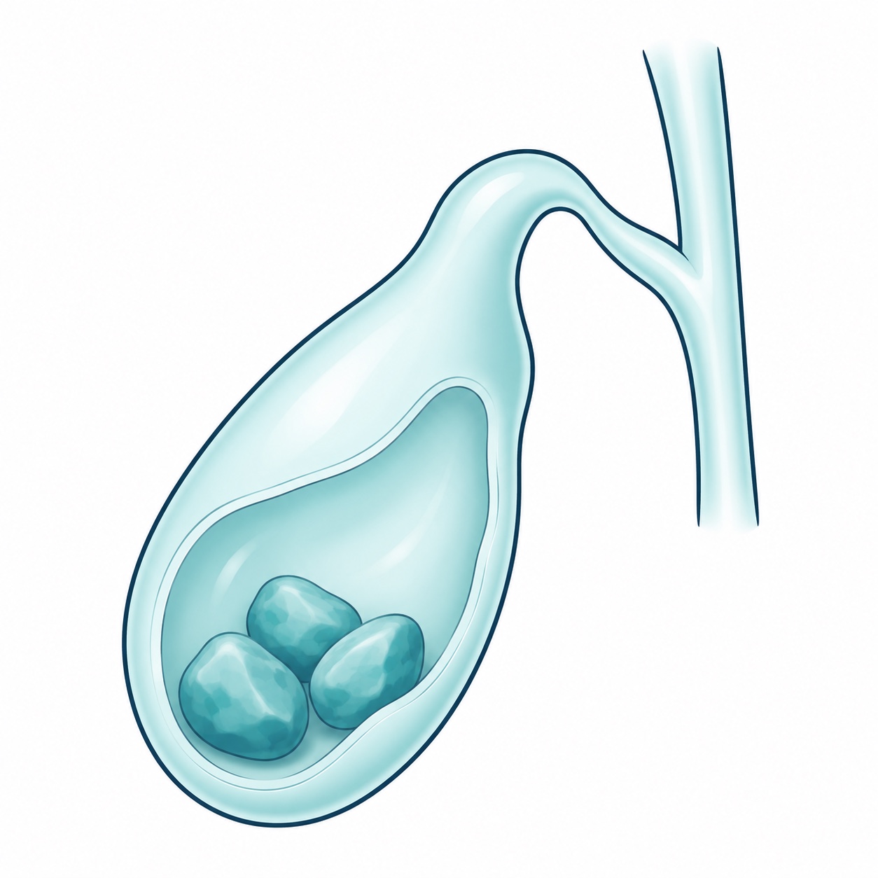
Illustration · Gallbladder with gallstones Removes the gallbladder when stones or other problems cause symptoms or complications. It may be performed laparoscopically or through open surgery, depending on the findings.
- Bile duct exploration
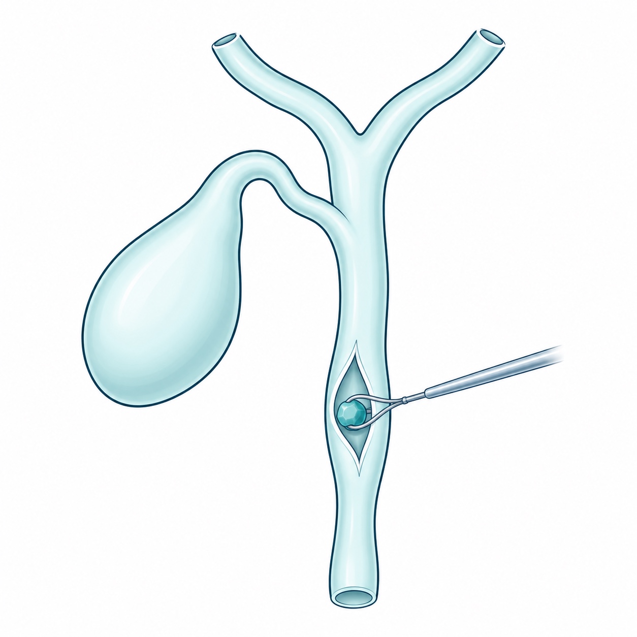
Illustration · Bile duct exploration Locates and removes stones from the ducts that carry bile to the intestine. It is considered based on tests, obstruction and the possibility of endoscopic treatment.
- Liver cyst surgery
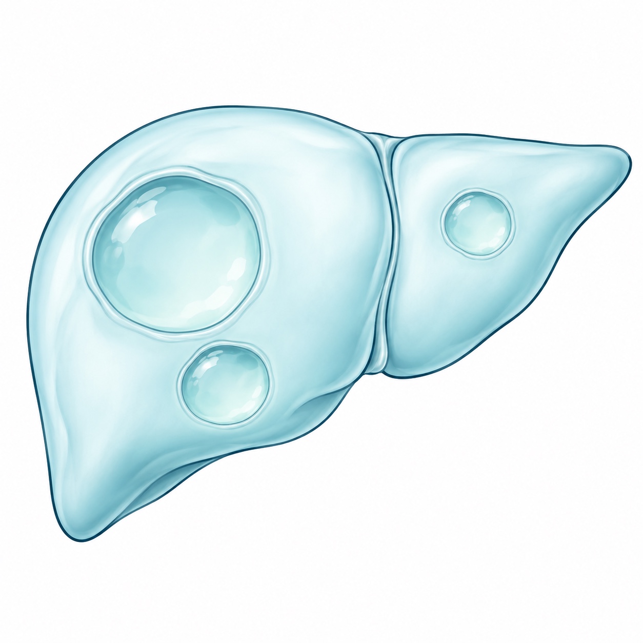
Illustration · Liver cysts Selected cysts that cause pain, compression or other complications may be drained and part of their wall removed. Treatment depends on the type of cyst.
Gastrointestinal and abdominal surgery
- Small intestine
Includes the duodenum, jejunum and ileum. Surgery may be needed for perforations, injuries, narrowing or diseased segments. Depending on the problem, the intestine is repaired or the affected portion is removed and the healthy ends joined with an anastomosis. As much intestine as possible is preserved to protect nutrient absorption.
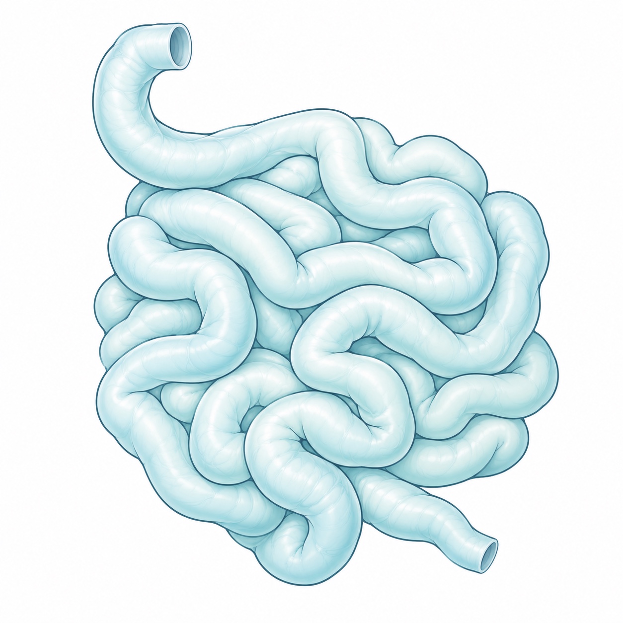
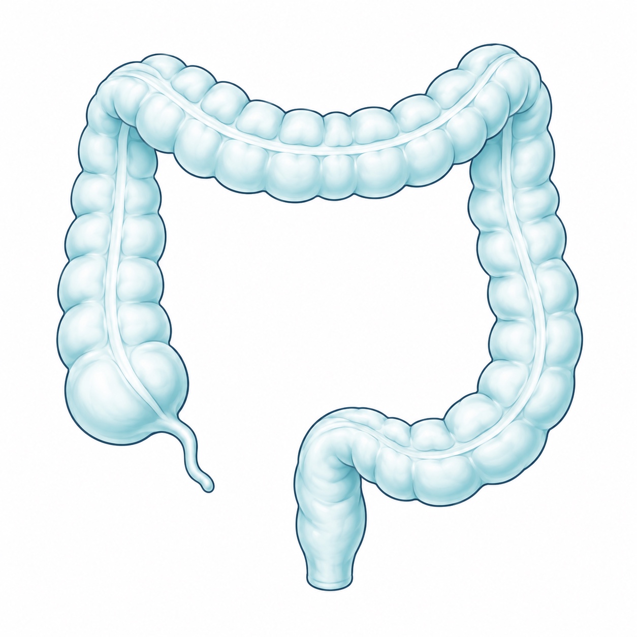
Small intestine · Illustration - Colon or large intestine
Includes the right, transverse, left and sigmoid colon. Some diseases require removal of part of the colon through a segmental colectomy or hemicolectomy. The extent depends on location and diagnosis. For tumors, evaluation includes biopsy and staging studies to plan treatment with the appropriate team.
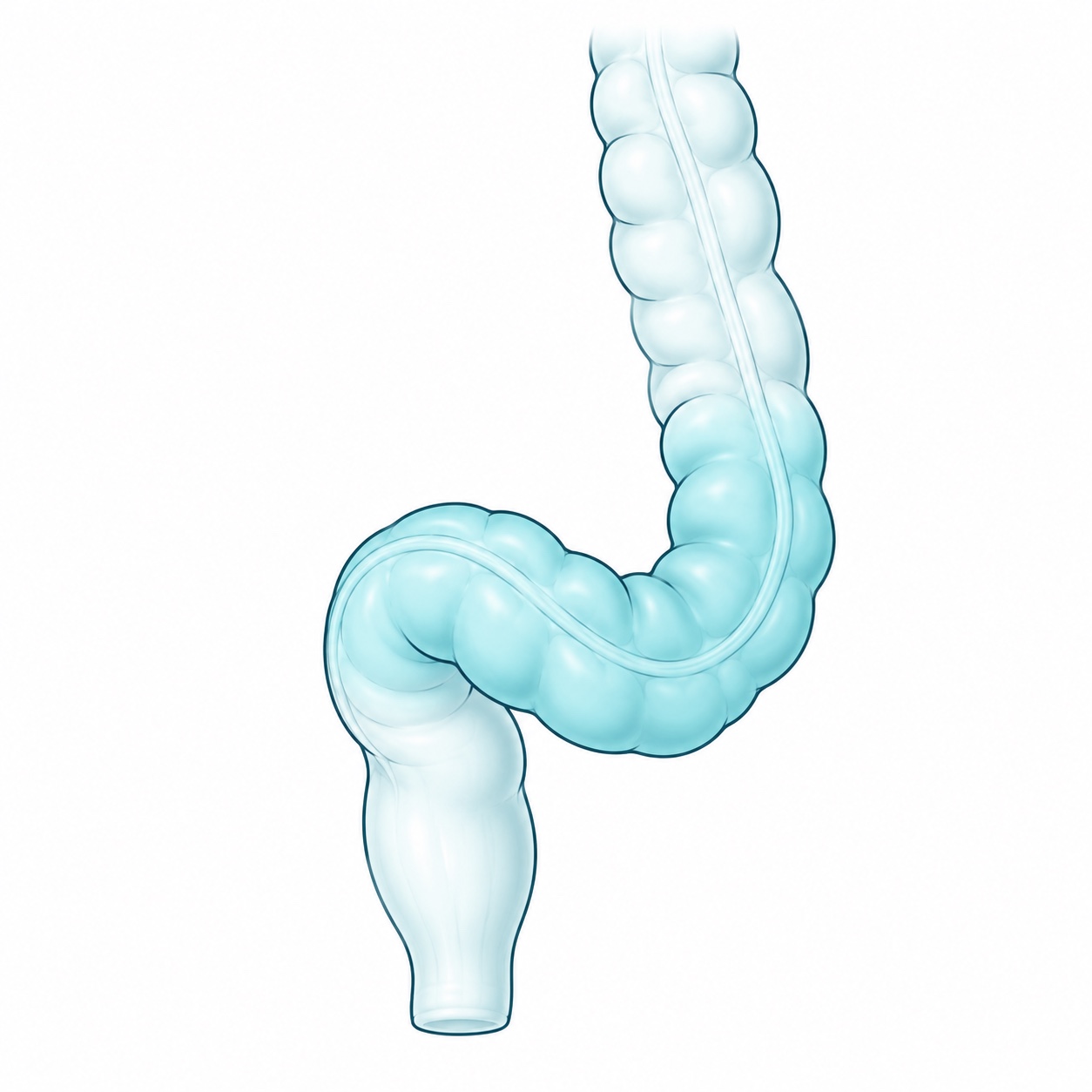
Colon or large intestine · Illustration - Sigmoid colon and sigmoidectomy
The sigmoid is the part of the colon that connects to the rectum. Sigmoidectomy removes this segment when indicated, such as for certain complications of diverticulitis or a tumor. If conditions permit, the colon is reconnected to the rectum; otherwise a colostomy may be needed.
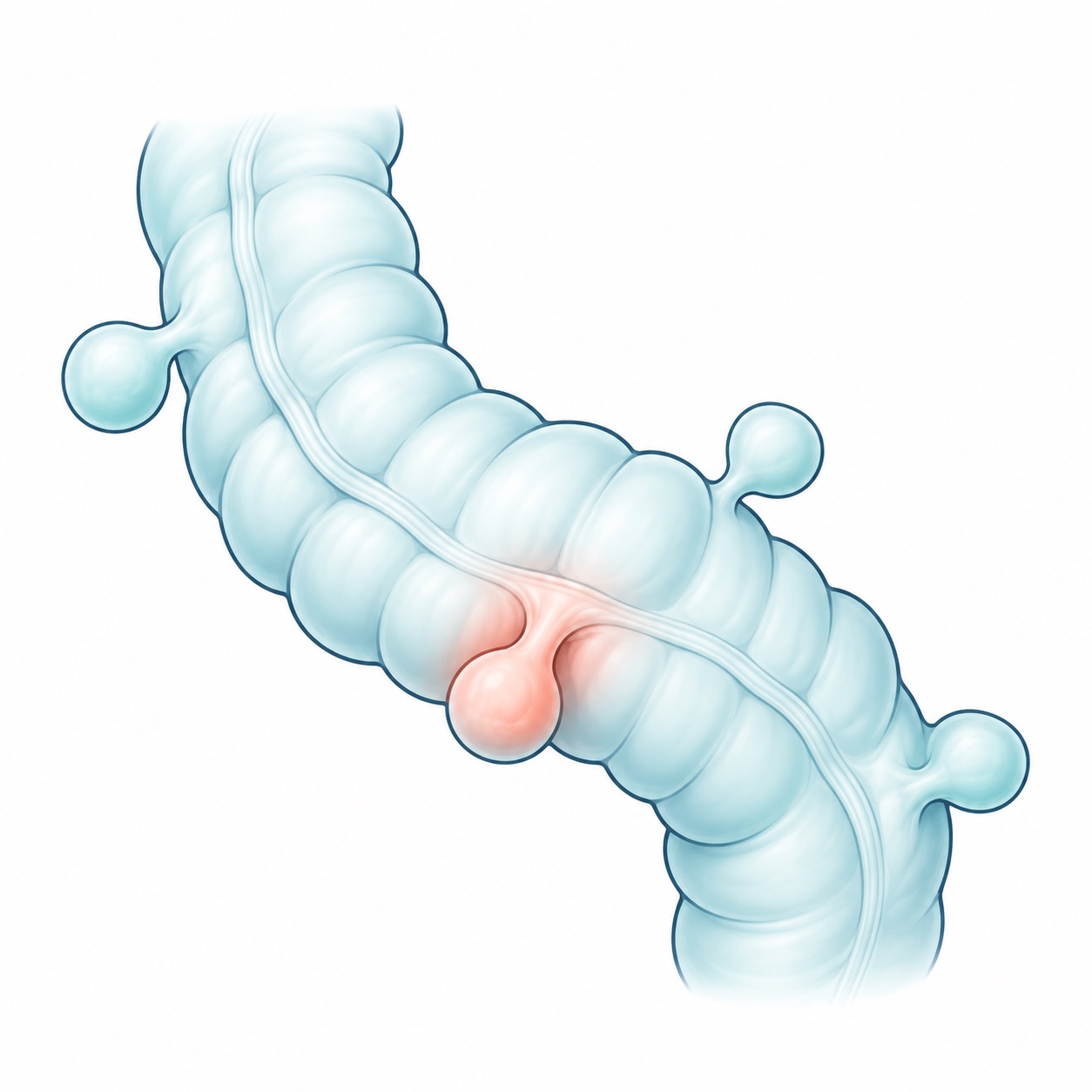
Sigmoid colon and rectum · Illustration - Diverticular disease and diverticulitis
Diverticula are small pouches in the colon wall. Inflammation of these pouches causes diverticulitis. Not every case requires surgery: the decision depends on the course of illness, complications and overall health. Perforation, fistula, obstruction or failure to respond to treatment may require removal of the affected segment.
Diverticula and diverticulitis · Illustration - Bowel obstruction and adhesions
An obstruction prevents intestinal contents from moving normally. Adhesions are internal scar bands, often related to previous operations, that can block the intestine. Some cases resolve with hospital treatment and monitoring; others require release of adhesions or removal of damaged tissue. Severe pain, vomiting, bloating and inability to pass gas require urgent care.
- Bowel resection and anastomosis
Resection removes diseased or damaged intestine. An anastomosis joins the ends to restore continuity. Whether this connection can be made depends on tissue blood supply, infection and overall condition. Risks include bleeding, infection, a leak at the connection and obstruction; follow-up helps detect complications.
- Ileostomy and colostomy
These are openings of the intestine onto the abdominal skin, called stomas, through which stool is collected in a bag. An ileostomy uses the small intestine and a colostomy uses the colon. They may be temporary or permanent depending on the disease and operation. Care includes guidance on the bag, skin, diet and hydration.
- Ostomy reversal and bowel reconnection
Restores passage of stool through the intestine in patients with a temporary ostomy. Recovery, anatomy and the safety of reconnection are assessed beforehand. Not all ostomies can be reversed, and there is no single timeline. Bowel movement frequency may temporarily change afterward.
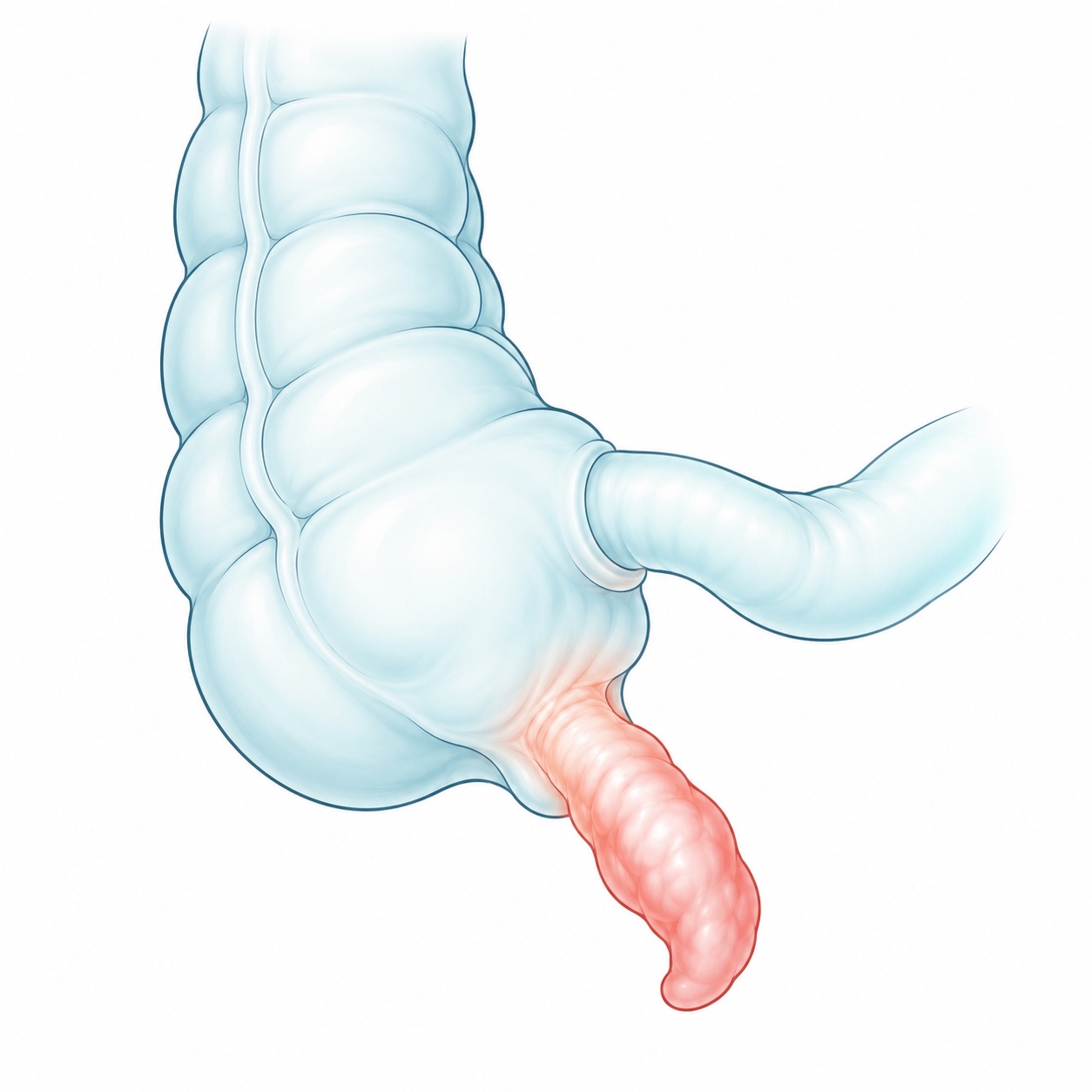
- Appendix and appendectomy
Appendectomy removes the appendix, usually for appendicitis. It can be laparoscopic or open, depending on the findings and the patient's condition. Perforation or abdominal infection may make treatment and recovery more complex. Early assessment of worsening abdominal pain helps identify its cause.
Appendix and appendectomy · Illustration - Stomach
Stomach surgery may repair a perforation or remove an area affected by a lesion requiring surgery. Depending on the diagnosis, endoscopy, biopsies and imaging help define the procedure. Gastric resection may require reconstruction of the digestive tract and dietary changes with nutritional follow-up.
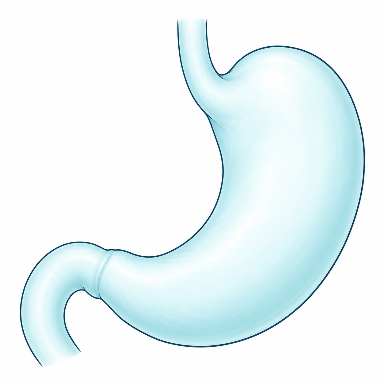
Stomach · Illustration - Pancreas
Surgical assessment focuses on selected pancreatic lesions, cysts or complications. Not every case of pancreatitis or pancreatic cyst requires surgery. Imaging and additional tests when appropriate guide the choice between observation, endoscopy and surgery. Complex procedures require a team and center with specific expertise.
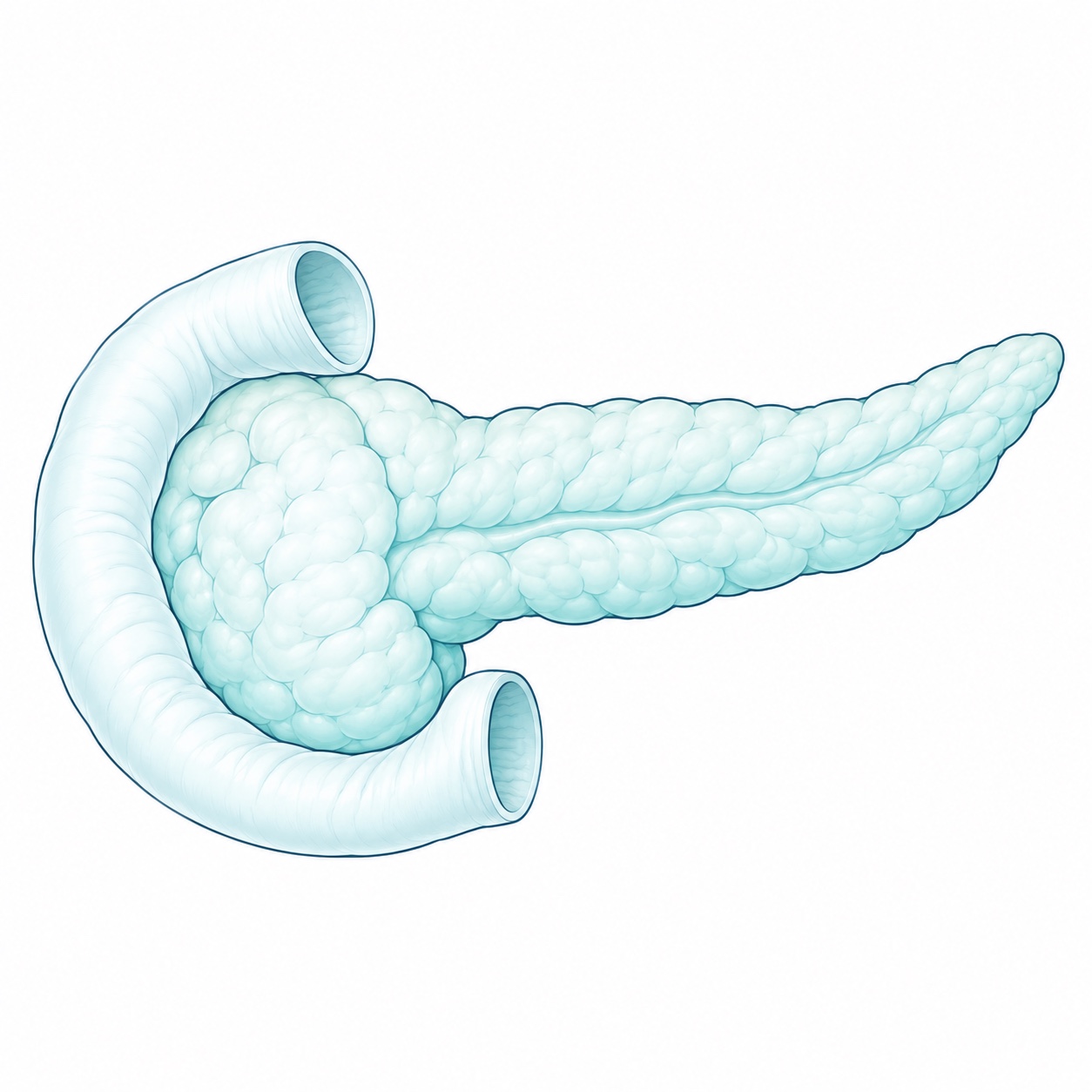
Pancreas · Illustration - Spleen and splenectomy
Some traumatic injuries, blood disorders or splenic lesions may require surgery. Splenectomy removes the spleen when indicated, while preservation is preferred when possible. Removal requires a vaccination and infection-prevention plan, plus instructions on when to seek medical care.
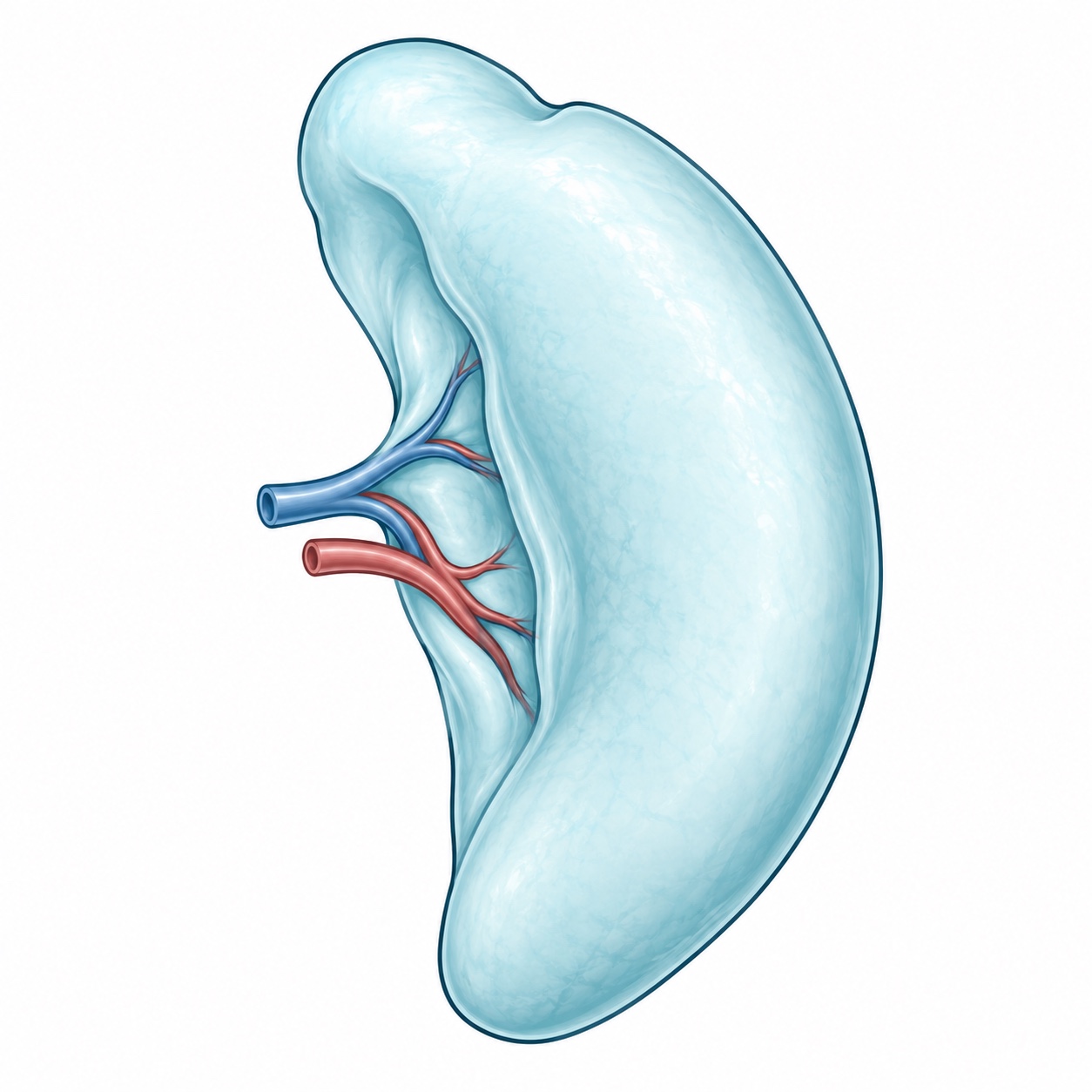
Spleen · Splenectomy · Illustration
The open or laparoscopic approach, hospital stay and return to activities are individualized. This information does not replace a medical assessment.
Patient information: diverticular disease, intestinal adhesions and ostomies (NIDDK).
Hernias
- Inguinal hernias
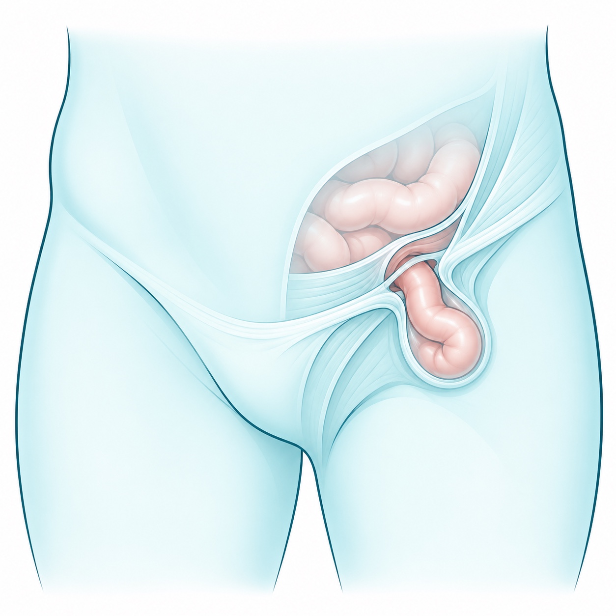
Inguinal hernias · Illustration These form in the groin when fat or part of the intestine protrudes through a weak area in the abdominal wall. They may cause a bulge, pressure or pain with exertion. Assessment helps determine whether observation in selected cases or surgery is appropriate. Repair may be open or laparoscopic, usually with mesh; symptoms, involvement of both sides and prior operations guide the choice.
- Femoral hernias
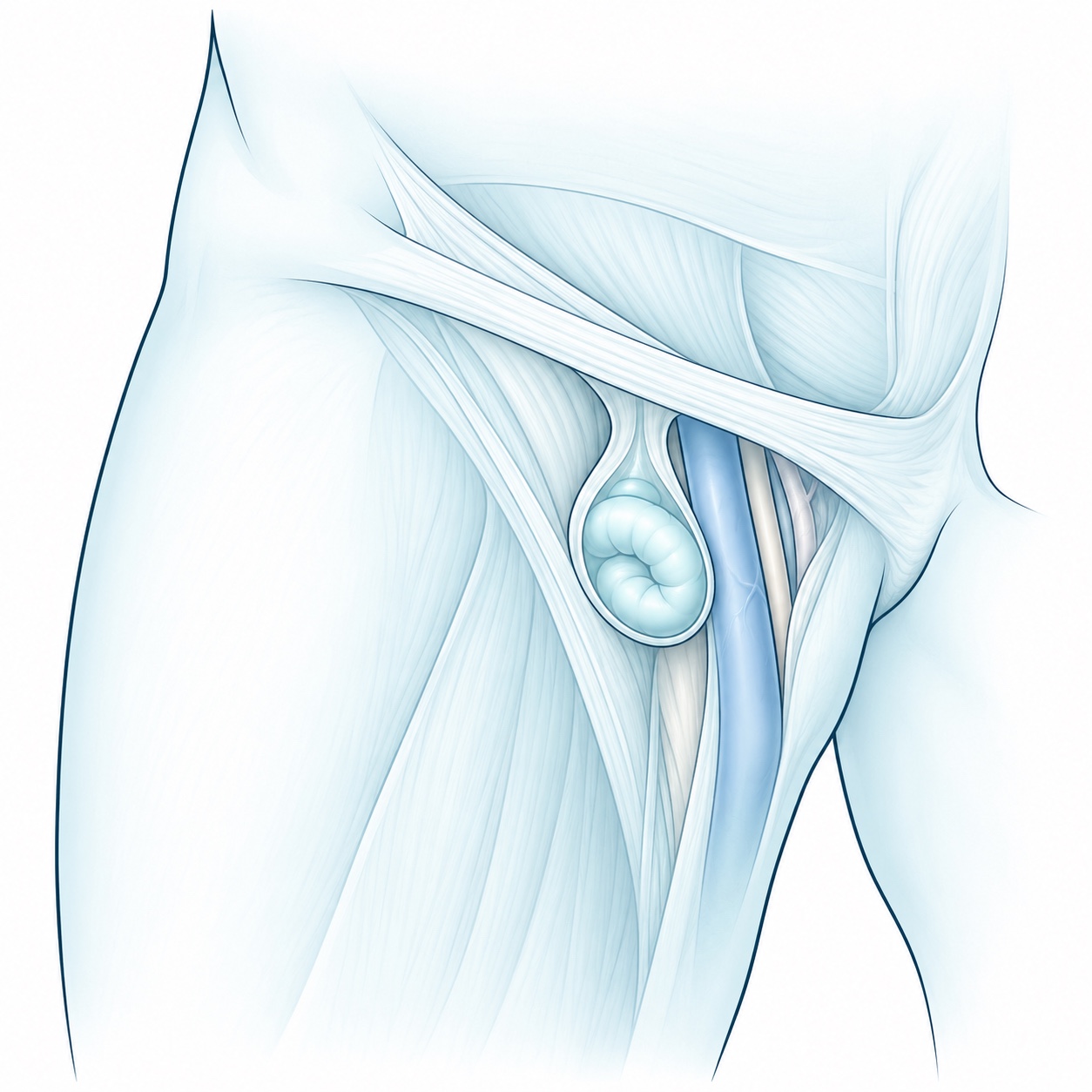
Femoral hernias · Illustration These occur below the groin in the upper thigh and are more common in women. The bulge may be small or difficult to identify. They carry a higher risk of trapped contents, so timely assessment is needed and surgery is often recommended. The open or laparoscopic approach depends on the findings and patient condition.
- Umbilical hernias
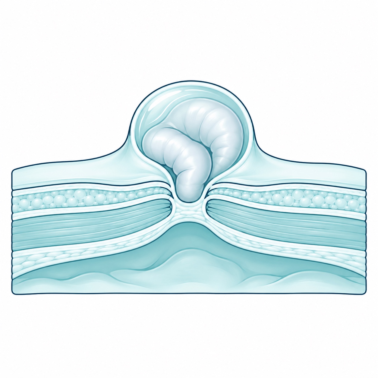
Umbilical hernias · Illustration These occur at the belly button, where fat or intestine protrudes through a defect in the abdominal wall. They can cause a bulge or discomfort when coughing, lifting or straining. In adults, repair closes the defect and may reinforce it with mesh depending on its size and the risk of recurrence. The timing of surgery is decided after reviewing symptoms and overall health.
- Epigastric hernias
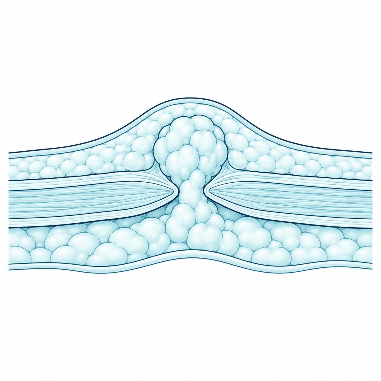
Epigastric hernias · Illustration These occur along the abdominal midline between the belly button and the lower end of the breastbone. They usually contain fat and may cause a small lump, tenderness or localized pain. Repair closes the opening; mesh use and the approach depend on size, symptoms and other defects.
- Ventral or abdominal wall hernias
Illustration · Abdominal wall defect These are defects in the abdominal wall through which fat or intestine may protrude. The term includes umbilical, epigastric and incisional hernias. Assessment considers location, size and effects on daily activities. Repair may be open or laparoscopic, with mesh reinforcement when appropriate; large hernias may require abdominal wall reconstruction.
- Incisional hernias
Illustration · Abdominal wall defect These develop at a previous surgical scar when the abdominal wall does not regain sufficient strength. They may enlarge and cause a bulge or discomfort. In selected cases, a CT scan helps plan repair. Treatment may involve closing the defect, placing mesh and using reconstruction techniques based on the extent and previous operations.
- Recurrent hernias
Illustration · Abdominal wall defect These return after a previous repair. Before recommending another operation, the previous technique, mesh location, studies and factors favoring recurrence are reviewed. Treatment is individualized. Other causes of postoperative pain are also evaluated: pain alone does not confirm recurrence.
- Hiatal and paraesophageal hernias
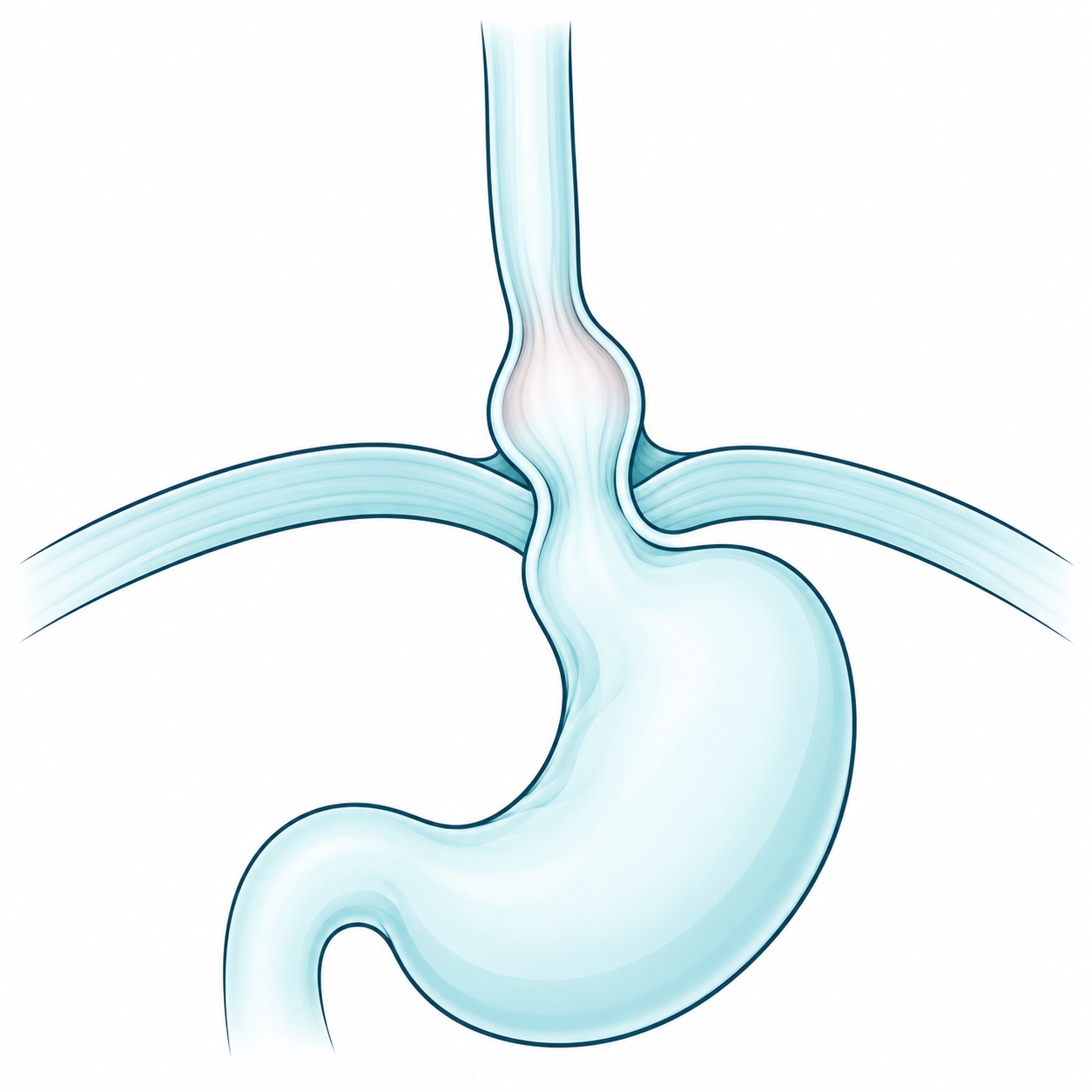
Hiatal hernia · Illustration 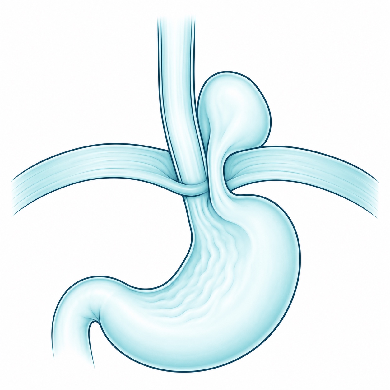
Paraesophageal hernia · Illustration These occur when part of the stomach moves into the chest through the opening in the diaphragm where the esophagus passes. They may be associated with reflux, regurgitation or difficulty swallowing, although some cause no symptoms. Not all require surgery. Repair returns the stomach to the abdomen and tightens the opening; fundoplication may be added to control reflux. The indication depends on hernia type, symptoms and tests.
When to seek emergency care: a very painful bulge that does not go down, vomiting, abdominal swelling or skin color changes require immediate care.
Patient information: American College of Surgeons and NIDDK.
Skin, soft tissues and thyroid
- Lipomas
Removal of fatty tissue masses, usually benign, when they cause discomfort, grow or need diagnostic confirmation.
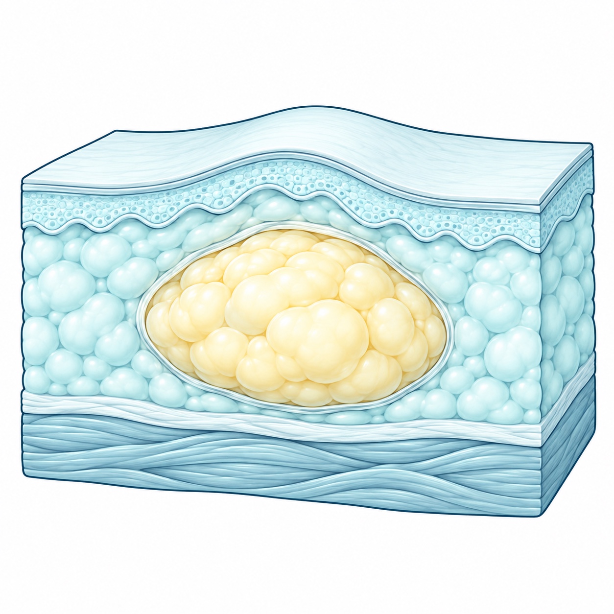
Lipoma · Lipoma removal · Illustration - Cysts and other benign skin and soft tissue tumors
Assessment and removal of selected lesions. Examination of the removed tissue can confirm its nature when appropriate.
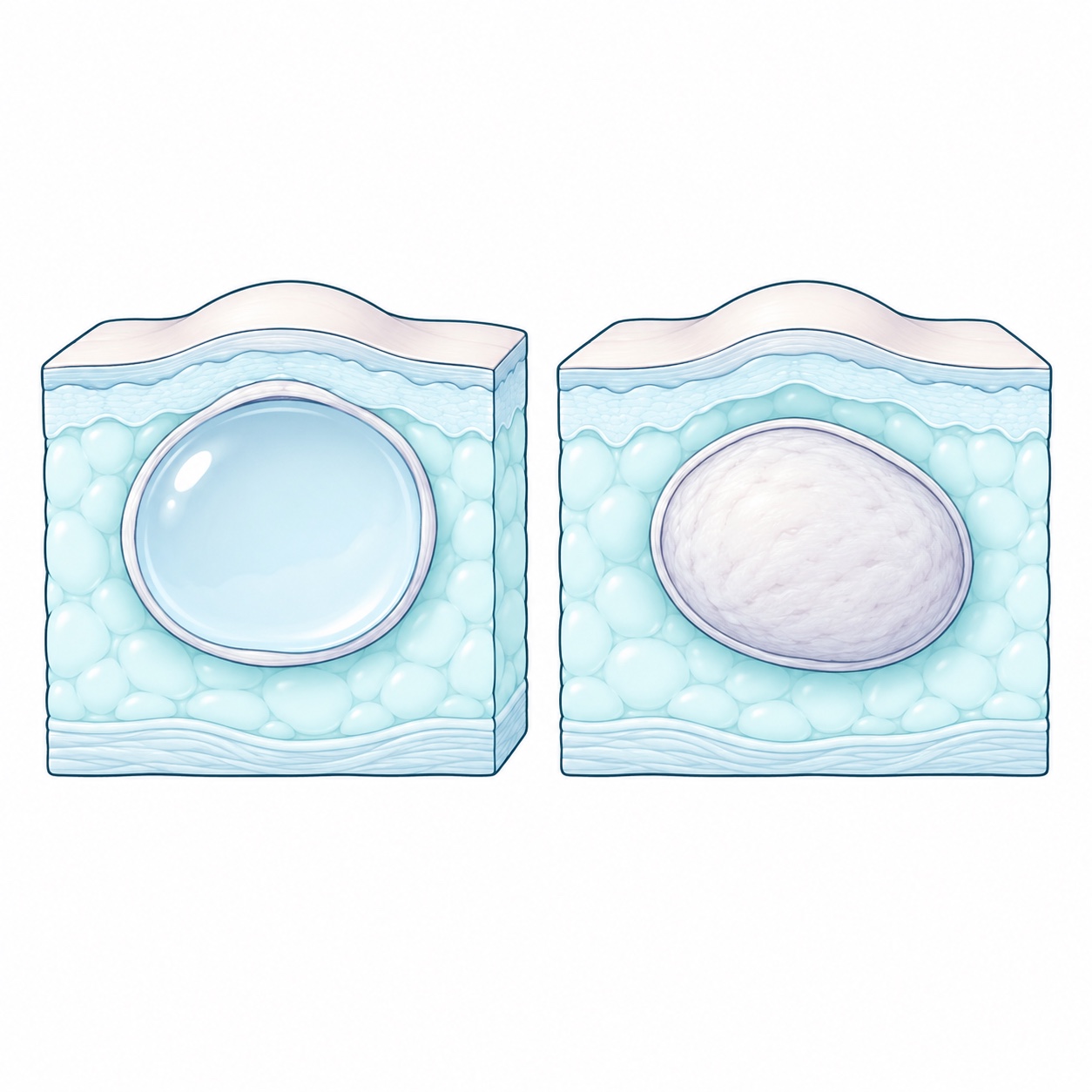
Skin and soft tissue cysts and tumors · Illustration - Thyroid surgical assessment
Evaluation of nodules and other abnormalities to determine whether surgery is needed. Decisions are based on ultrasound, thyroid function and biopsy when indicated.
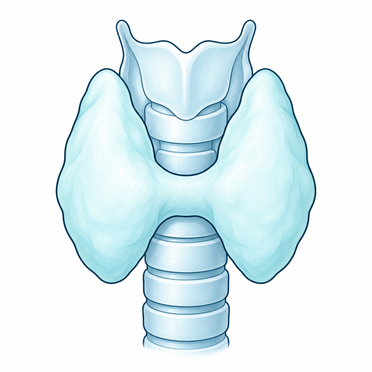
Thyroid · Thyroidectomy · Illustration
Bariatric and metabolic healthSurgery, medication and combined options.
Body mass index calculator
Enter your weight and height to calculate your BMI.
Guidance for adults. BMI alone does not determine which treatment you need; a medical assessment also considers your health and history.
Surgical procedures
- Sleeve gastrectomy
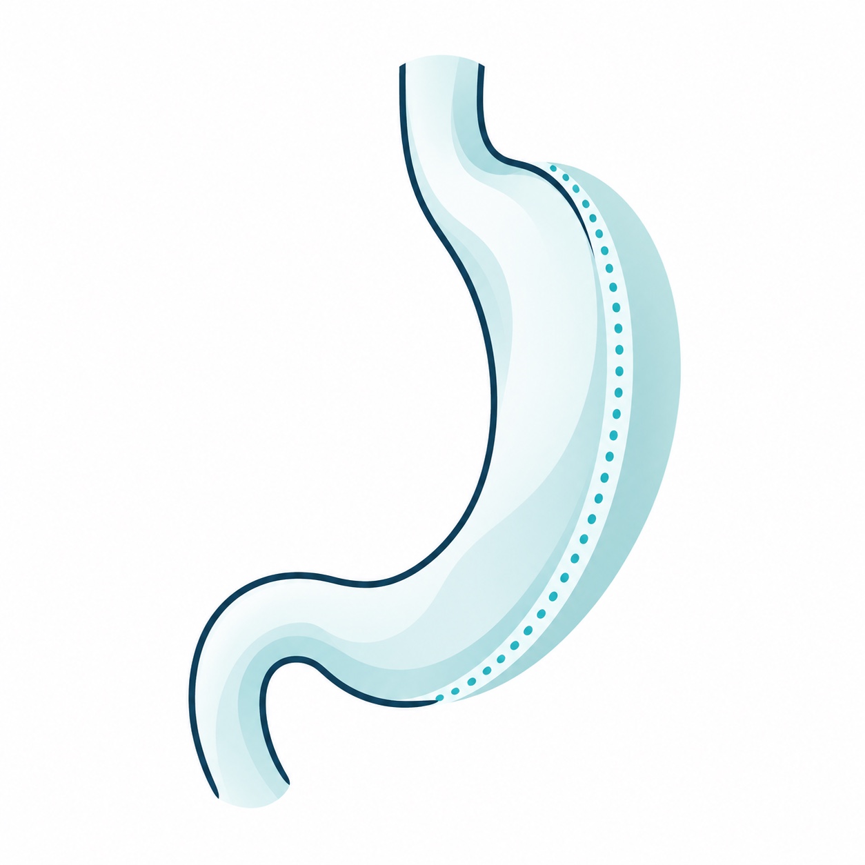
Illustration · Sleeve gastrectomy Removes part of the stomach to form a narrower tube. It changes stomach capacity and appetite-related signals; nutritional follow-up and reflux monitoring are required.
- Gastric bypass
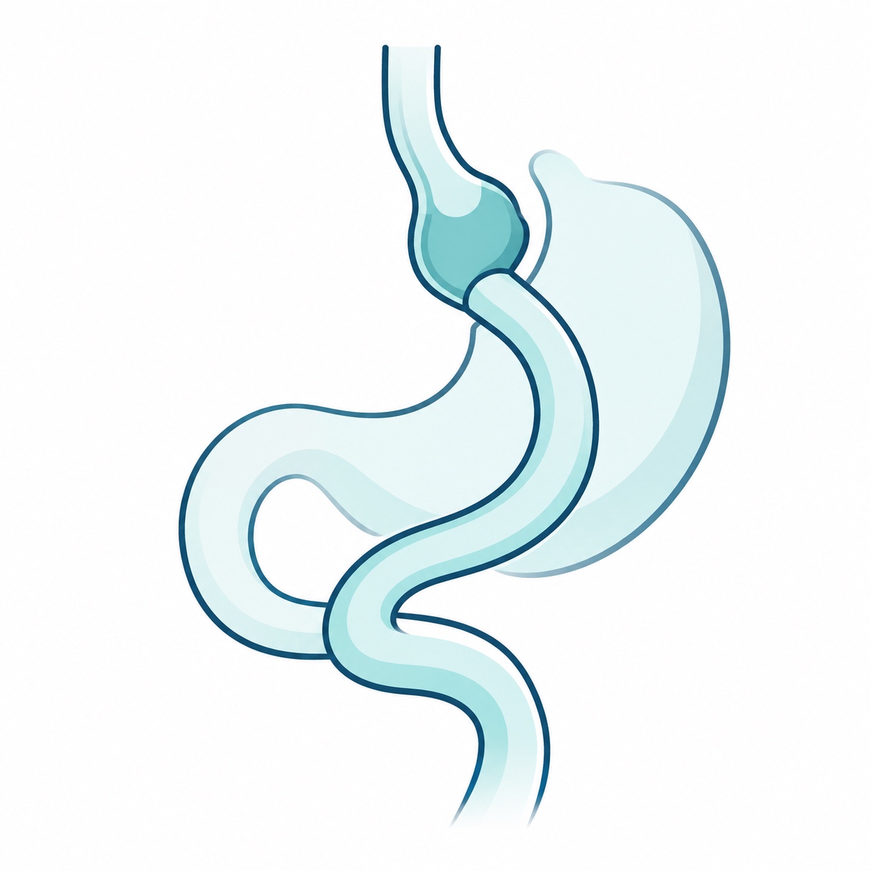
Illustration · Gastric bypass Creates a small stomach pouch and changes the route food takes through the intestine. Selection considers metabolic health, reflux and the need for long-term supplements.
- Adjustable gastric band
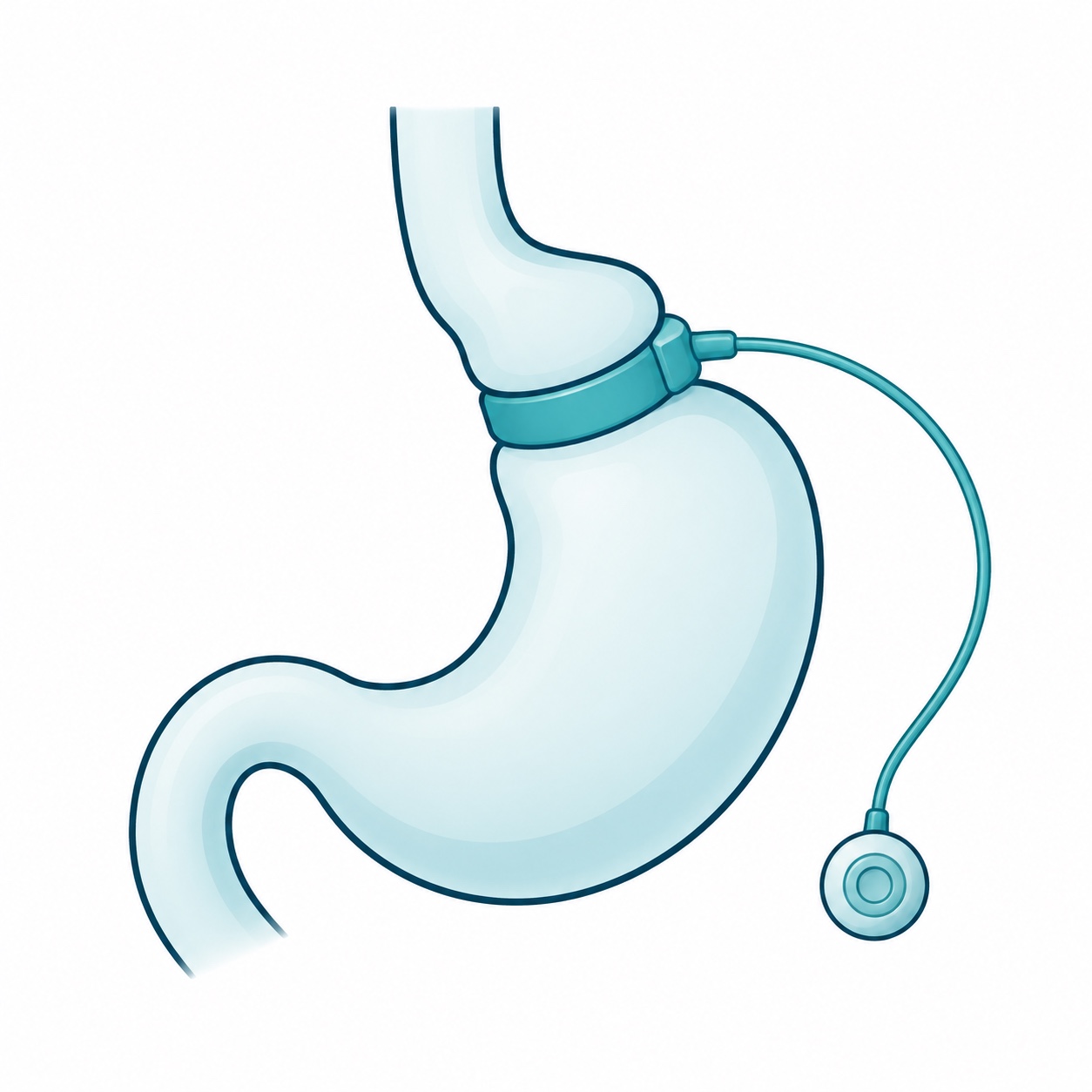
Illustration · Adjustable gastric band Places a band around the upper stomach. Adjustments and follow-up are needed; removal or conversion may be considered for symptoms or insufficient results.
- Duodenal switch
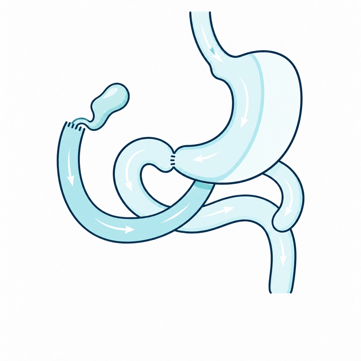
Illustration · Duodenal switch Combines sleeve gastrectomy with a major change in intestinal routing. Close nutritional monitoring, supplements and lifelong blood tests are essential.
- SADI-S
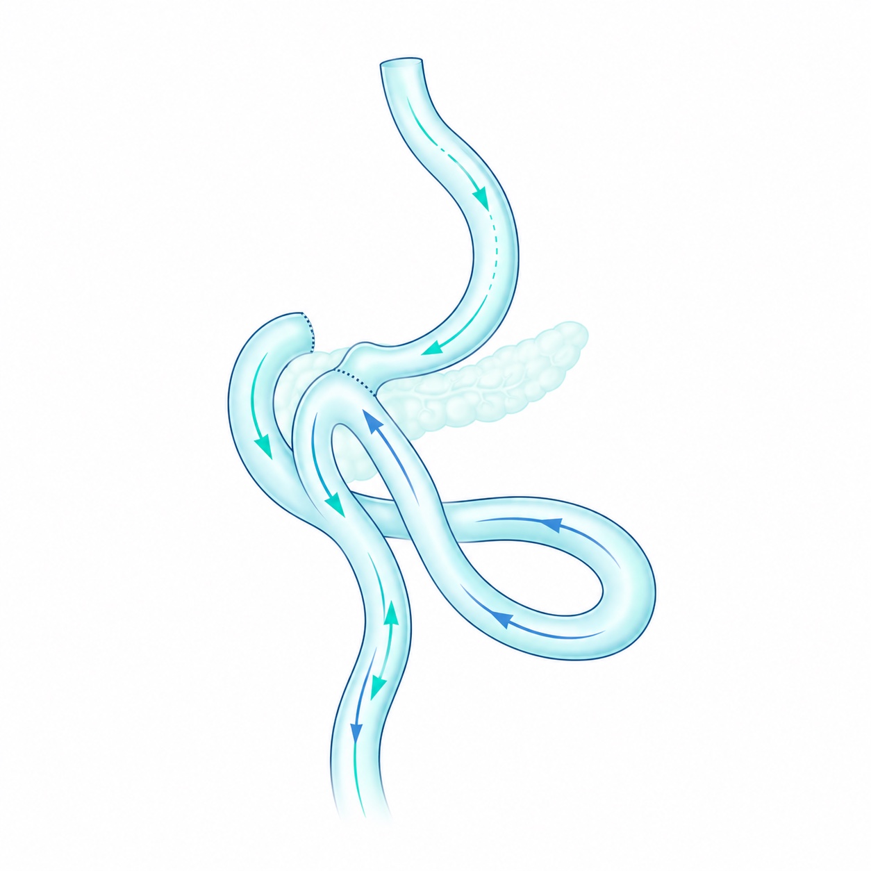
Illustration · SADI-S Combines sleeve gastrectomy with a connection between the duodenum and ileum. It may be considered as an initial or revision operation in selected patients and requires lifelong nutritional follow-up.
- SASI or intestinal bipartition
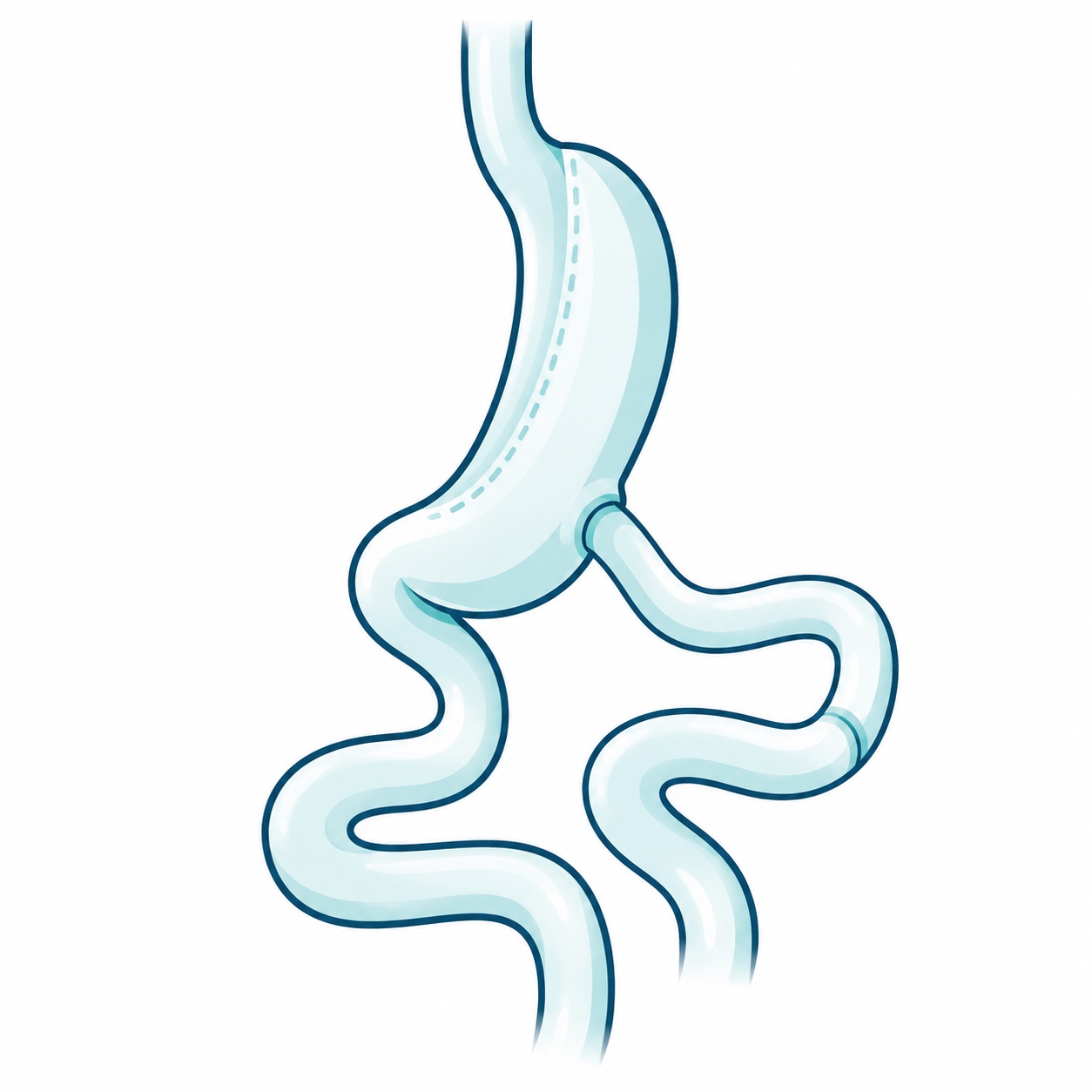
Illustration · SASI or intestinal bipartition Combines sleeve gastrectomy with an additional stomach-to-intestine connection while preserving passage through the duodenum. The technique and nutritional implications are discussed during consultation.
- Revision surgery
Modifies a previous bariatric operation for complications, reflux or insufficient response. Anatomy, tests and progress are reviewed first to identify alternatives.
Medication treatment
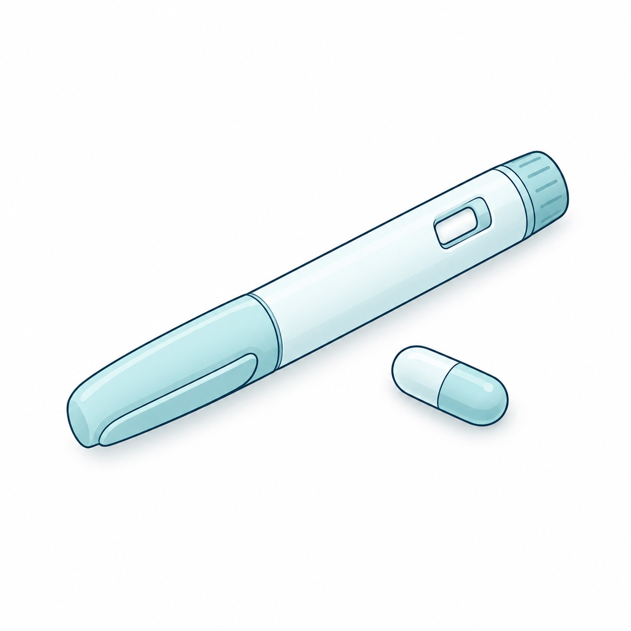
Oral and injectable options are available. Injectable medicines with a weight-management indication documented by COFEPRIS include liraglutide (Saxenda) and semaglutide (Wegovy). Selection and availability must be confirmed during consultation; other medicines registered for diabetes do not necessarily have an obesity indication.
Combined treatment
In selected cases, lifestyle changes, medication, endoscopy and surgery can be combined. The sequence and follow-up are tailored to each person.
Bariatric endoscopyGastric balloon, endoscopic sleeve and outlet reduction.
These procedures use an endoscope introduced through the mouth. Choosing a procedure requires individual assessment.
Intragastric balloon
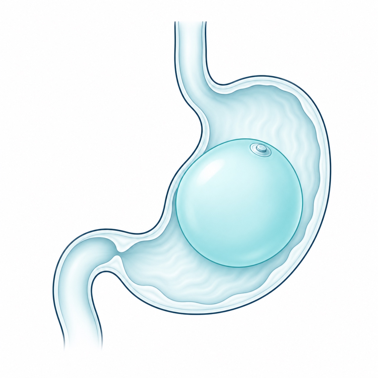
A temporary device that occupies space in the stomach and promotes fullness. It should be combined with a nutrition and follow-up program. Duration, placement and removal depend on the model used.
Endoscopic sleeve gastroplasty (ESG)
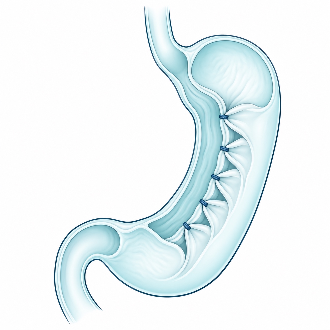
Endoscopically placed sutures reduce the stomach's internal space without removing part of it. Lifestyle changes and follow-up are required; outcomes and metabolic effects differ from surgical sleeve gastrectomy.
Transoral outlet reduction after bypass (TORe)
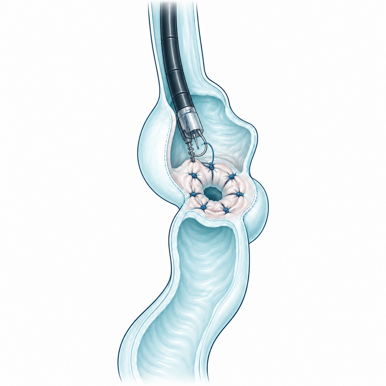
In selected patients with previous bypass, endoscopy reduces an enlarged connection between the stomach pouch and intestine. It may help with weight regain or certain dumping symptoms after the cause has been evaluated.
Esophagus and refluxAchalasia, hiatal hernia and difficulty swallowing.
Esophageal assessment and surgery
Similar symptoms can have different causes. Before proposing surgery, the necessary tests are reviewed to establish the diagnosis.
Achalasia
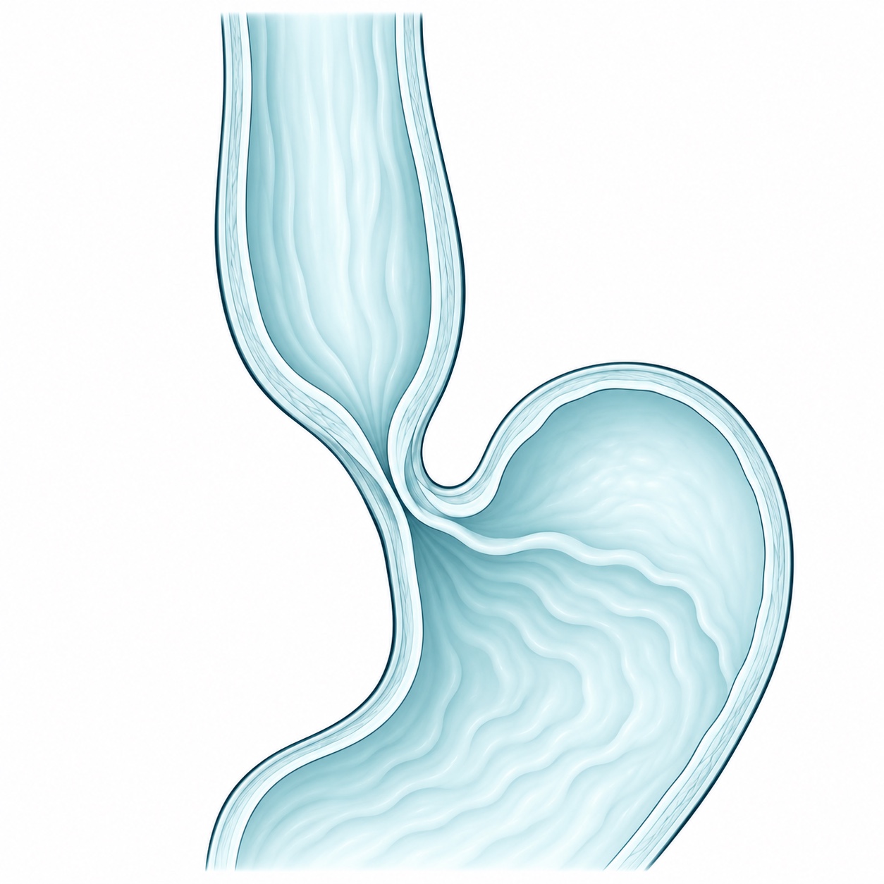
Assessment using endoscopy, esophagram and manometry. When appropriate, surgical treatment includes Heller myotomy and Dor fundoplication.
Reflux and hiatal hernia
Read about reflux and hiatal hernia
Evaluation of gastroesophageal reflux and hiatal hernia. Repair and fundoplication, including the Nissen technique, are considered based on the findings.
Difficulty swallowing
Evaluation of dysphagia to determine whether it is related to a motility disorder, obstruction or another cause.
Symptoms after previous surgery
Assessment of reflux or difficulty swallowing after antireflux or bariatric surgery to determine the next step.
Laparoscopic surgeryThe technique and its uses in different operations.
Laparoscopy uses small abdominal incisions, a camera and surgical instruments. It differs from digestive endoscopy. Each procedure description explains whether this approach may be an option.
Gastrointestinal
Gallbladder surgery, selected hernias, reflux and digestive procedures as appropriate.
Bariatric and metabolic surgery
Sleeve gastrectomy, bypass and other procedures selected after a comprehensive assessment.
Adrenal glands
Adrenalectomy removes an adrenal gland when a lesion or its hormone production requires it. Hormones and imaging are evaluated before surgery; lesion size and characteristics determine the approach.
Spleen
Splenectomy removes the spleen for selected blood disorders or injuries. The approach depends on diagnosis and organ size. Vaccinations and infection-prevention measures are reviewed when removal is planned.